Raman Spectroscopy
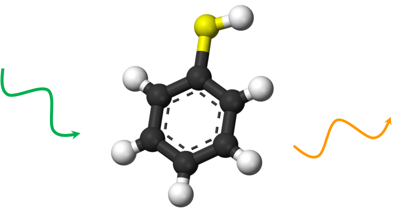
Raman Spectroscopy is a non-destructive technique which provides structural and electronic information from materials. It allows the identification of substances and the calculation of their concentration in samples. Raman spectroscopy is based on the interaction of light with chemical bonds within the material and therefore does not require stains or labels.
In combination with an imaging system, Raman spectroscopy can be used to generate maps based on the sample’s Raman spectrum. These images show distribution of individual chemical components, variation in crystallinity, stress, doping, etc.

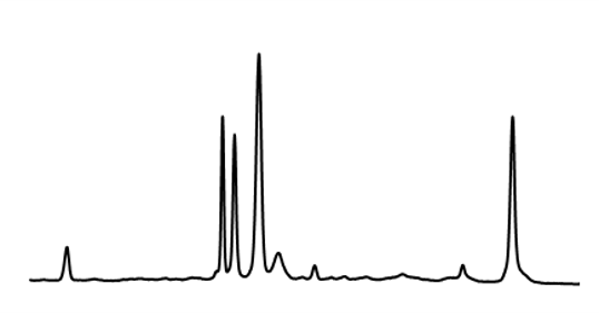
Raman spectroscopy provides unique vibrational fingerprints by which molecules can be identified

Surface-enhanced Raman spectroscopy or surface-enhanced Raman scattering (SERS)
One drawback of Raman spectroscopy is the weakness of the Raman scattering intensity. However, this situation can be easily improved by placing the Raman emitter in the proximity of a noble metal surface, where signal enhancements of up to 10^11 allow for the detection of Raman spectra at the single molecule level.
There are two mechanisms behind this Surface-Enhanced Raman Scattering (SERS): The so-called chemical enhancement, and the electromagnetic field enhancement. The chemical enhancement can be explained by the formation of charge-transfer complexes.
The electromagnetic field enhancement is the dominant mechanism and is composed of three different contributions: The antenna effect, the lightning rod effect and the presence of localized surface plasmons. Localized surface plasmons are collective electron oscillations that are controlled by the shape and the material composition of the nanostructure supporting them and might result in particularly strong local fields in the optical regime. The strongest signal enhancements are generated at the electromagnetic field hot spots created at the interstitial sites between nanoparticle aggregates.
Equipment

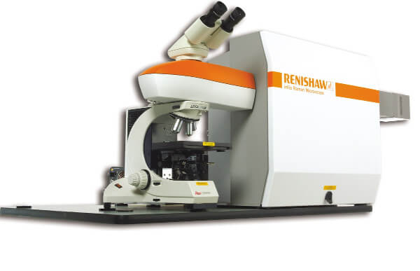
InVia Raman microscope from Renishaw plc.


